The Imaging Shared Resource is a core facility with the primary goal of providing exceptional microscopy and imaging services, as well as individual access to a variety of state-of-the-art imaging resources for Wistar users and members of the local research community. The imaging systems have been designed to be extremely flexible in order to reflect a broad range of challenging scientific questions and specimens. These systems allow researchers to determine how the temporal and spatial organization of regulatory events within cells, tissues and organisms impact both normal and pathological processes.
All services of the Imaging Shared Resource are coordinated through the main laboratory in suite 287, although some equipment is placed in remote locations. The Imaging Facility staff can provide all listed services or researchers may be authorized to work with the equipment unassisted. Interested users must be trained on any equipment they wish to use before unassisted operation of the equipment is permitted.
We Provide:
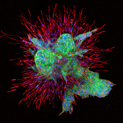
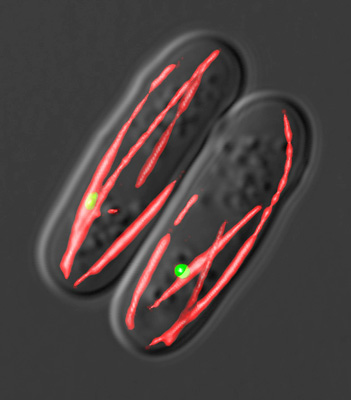
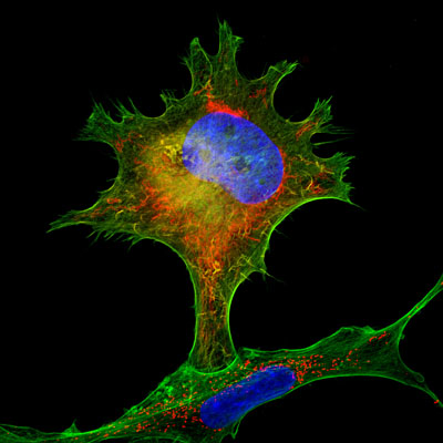
CONFOCAL EQUIPMENT Current major equipment includes both confocal and widefield systems, including our Leica Stellaris 8 3X TauSTED system for fixed or live experiments using traditional spectral confocal techniques from 440-790nm, FLIM (fluorescence Lifetime Imaging) and super-resolution STED. We also have a Leica TCS SP8 X white light confocal system with stage-top environmental control (including hypoxia), and our TCS SP8 MP intravital 2-photon microscope built on a fixed-stage upright system. All confocals include resonant scanning and sensitive HyD detectors and provide excellent channel separation.
WIDEFIELD AND OTHER SYSTEMS Widefield systems include automated Nikon NiE and manual 80i upright microscopes and automated Nikon TiE and manual TE2000 inverted microscopes, and a Leica MICA microhub, all equipped for brightfield, fluorescence, color and monochrome image capture. A semi-automated live-cell time lapse system based on a Nikon TE300 inverted microscope is also available. Whole Slide Imaging (WSI) is available as a service using our Hamamatsu Nanozoomer S60 microscope Nikon SMZ1500 and 800 stereomicroscopes. The NiE upright, TiE and MICA microhub inverted systems are fully automated for capture and analysis with Nikon AI software and Leica LAS-X. Whole Slide Imaging, as well as full plate scanning is available.
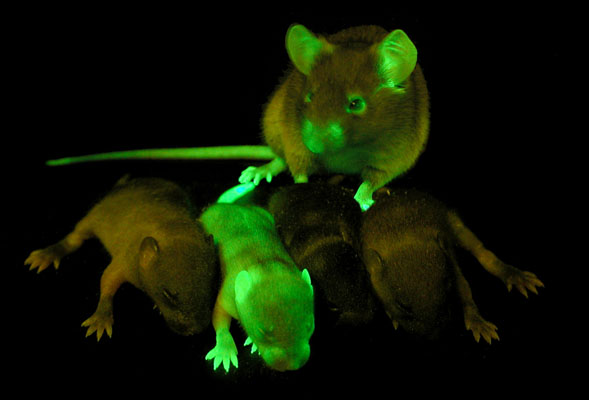
SMALL ANIMAL IMAGING AND RADIOTHERAPY The Facility provides a series of instrumentation housed inside the Wistar Animal Barrier Facility taking advantage of a variety of whole-body 3D imaging modalities for mice. Systems include a Revvity SpectrumCT providing whole body luminescence, fluorescence, and microCT imaging and a Revvity Vega small animal ultrasound system, including a special set-up for image-guided injection. Outside of the barrier, a SARRP (Small Animal Radiation Research Platform) is available for whole-body and targeted radiotherapy of mice.
PHOTOGRAPHIC SERVICES Traditional photographic services are available in the main lab for a wide variety of subjects, including whole dishes or plates, special low magnification (photomacrography) applications, live mice, gross specimen photography, location and group photography.
TRAINING, ASSISTANCE & ANALYSIS Users of the Imaging Facility may be trained for unassisted use of most core assets, or they may take advantage of the expertise of the facility staff to request assisted service with the staff performing the imaging and providing analysis instead. The Imaging staff also provides assistance to researchers with other aspects of their experimental design: ideal approaches to specimen documentation are often unique to the experiment, and the staff can help determine the most effective imaging protocols to answer a particular question. Imaging Facility staff are also available to help investigators get the most out of their own microscopes. Training and basic maintenance (including cleaning, alignment and changing bulbs) are available on request. Customized image analysis with Nikon AI software, Photoshop training, creative imaging for journal covers, and guidance on digital imaging ethics help to round out the services available from the Facility.
Additional Services Include:
Widefield Microscopy - Conventional microscopy utilizing standard techniques in brightfield, darkfield, fluorescence, phase contrast and differential interference contrast is available on a variety of instruments in the main Core Facility suite 287. Upright, inverted and stereomicroscopes capable of low to high magnification documentation are available with individual image capture workstations networked to the Wistar servers.
Live-Cell Time-Lapse Microscopy – Using the Nikon TiE and TE300 inverted microscopes in suite 287, we can run multiple time-lapse studies of cells grown in culture. With the incubation chamber, XY stage and Z axis controller, we can track multiple areas in separate wells at the same time in multiple fluorescent channels. Stable imaging for up to 72 hours can be accommodated with longer sequences possible. Cells may be grown in 6 well plates or 35mm dishes as standard, but we can accommodate other vessels as needed.
Confocal Microscopy - The Imaging facility maintains Three Leica confocal systems including the newest Stellaris 8 3X TauSTED microscope, an SP8 WLL system and an SP8 intravital, 2-photon microscope. These systems are higher resolution and allow true 3D (Z-stack) capture in multiple channels, and multiple locations (6D imaging). The Stellaris includes FALCON fluorescence Lifetime Imaging as well as STED super-resolution in 3 channels. The integrated white-light laser accomodates dyes with excitations ranging from 440 to 790nm. The system is housed in a full OKO labs environmental chamber which can accomodate any sample holder for live-cell imaging The SP8 WLL confocal uses a laser that ranges from 460 - 660nm excitation and includes a 3-gas mixer for live-cell experiments that need hypoxic conditions. The SP8 MP intravital microscope can use 2-photon imaging techniques on live animals, explant tissues or fixed samples
Whole Slide Imaging (WSI) - The facility offers a whole slide imaging service in both brightfield and fluorescence using a Hammamatsu Nanozoomer S60 imager. Each slide is scanned at an equivalent 40X resolution (brightfield) or 20X (fluorescence) for a fixed price, regardless of the size of the area. The system can accomodate up to 60 slides in a single run and up to 5 fluorescent channels can be captured. Once scanned, the images are uploaded to a cloud-based storage and visualization platform called Concentriq for protected access from any device. HIgher resolution and or special case samples can also be imaged using our Nikon NiE and TiE systems. These microscopes have automated XYZ stages and can be configured for unattended imaging, automated image stitching and full or semi-automated analysis. Variably sized sample containers can be accommodated in the TiE for customized imaging.
Image Post Production and Analysis - Image capture is just the first part of acquiring high quality images. Whether taken with Core equipment or other laboratory microscopes, the next step in optimizing images involves image processing and analysis. The facility staff is available to train users both on the intricacies of the image capture software as well as popular third party image processing programs such as Nikon Elements AR, Leica LAS-X, SVI Huygens Professional, QuPath, FIJI and Adobe Photoshop. We have the ability to design custom software macros to help objectively quantify your results and process large data sets. We can run the analysis on your images or show you how to do it yourself.
Macro, Specimen, Small Animal and other Specialty Image Capture - We provide additional expertise in a variety of specialized scientific imaging situations such as photomacrography, small animal and gross specimen photography, gel and blot documentation, ultraviolet imaging as well as fluorescence in larger subjects, like whole mice. Additionally, creative imaging and graphic design for journal covers and public relations uses can provide proven exposure to the research and to the Institute as a whole. These specialized techniques can be provided as a direct service or for training to those interested in long-term applications. Special set-ups to capture 1-shot images of large histology sections for analysis are available. Our equipment and services are flexible enough to meet almost any need.
Group photography of your lab is also available at your convenience.
Nan Zhang, Ph.D.
Scientific Director
nzhang@wistar.org
Pavan Vedula, Ph.D.
Managing Director
pvedula@wistar.org
Frederick Keeney
Assistant Managing Director
fkeeney@wistar.org
Vic(Fengchong) Kong
Co-Managing Director
fkong@wistar.org
Sam(Shang) Wu
Research Assistant II
swu1@wistar.org
| Hours | Location |
|
8:30am - 5pm Monday - Friday
|
The Wistar Institute |
| Name | Role | Phone | Location | |
|---|---|---|---|---|
| Pavan Vedula |
Managing Director
|
(973) 366 6222
|
pvedula@wistar.org
|
287
|